










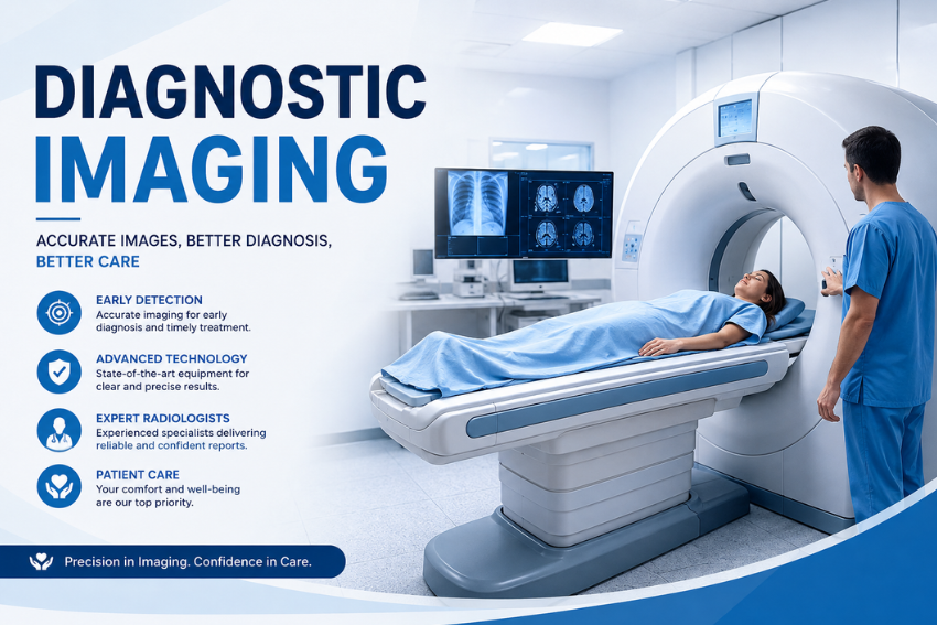
Diagnostic imaging is a key discipline that supports the practices of healthcare workers of various specialties (including physicians in numerous fields such as radiologists, cardiologists, oncologists, etc.). Such imaging techniques allow visualizing structures inside the body without only rely on direct or indirect observation through the external surface and through probing techniques. Based on the technological principles underlying particular techniques, physicians are often able to visualize bones, solid and hollow organs, soft tissues, circulatory system and cellular processes, metabolic activity, etc.
The global dimension of diagnostic imaging is enormous. According to estimations from the World Health Organization (WHO), annually at least more than 4.2 billion of diagnostic radiography procedures have been carried out worldwide. Also, nuclear medicine procedures are annually estimated to be about 40 million for a total usage (global utilization rates) of around 20 or 15 procedures or so per 1000 population, suggesting a very high diagnostic, screening, guiding, therapeutic planning, post therapeutic follow-up rate.
Conventional methods such as X-ray technology, Computed Tomography (CT), Magnetic Resonance Imaging (MRI), Ultrasound, Nuclear Medicine, Mammography, Fluoroscopy are some of the known diagnostic imaging devices. Recently artificial intelligence (AI), advanced detectors, fully automatic reconstruction, quantitative imaging are some examples which has been transforming medical imaging practice.
What Is Diagnostic Imaging?
Imaging is the technique and procedure of creating visual representations of the interior of a body for clinical analysis and medical intervention. Imaging can detect or record normal structures, abnormal pathology, or to locate a pathologic lesion or abnormality. It is one of the diagnostic tools available to evaluate symptoms, diagnose a medical condition, investigate symptoms or injury, plan medical, surgical or radiation treatments, and monitor the efficacy of treatment.
Medical imaging employs a variety of different physical principles. It can be performed by X-ray, CT (electromagnetic radiation), MRI (magnets and radiofrequency radiation), ultrasound (ultrasound waves) and nuclear medicine (radioactive tracers).
There are a number of other uses for diagnostic imaging. For example, a doctor may order an X-ray to look for a suspected broken limb, a CT to look for internal injury, an MRI to look at internal soft tissue structures or an ultrasound to examine blood flow.
Imaging can also be used for screening as well as for the monitoring of treatment. A screening mammogram, for instance, can determine if breast cancer is present. Additional CT scans, MRI, PET scans, etc., might be used multiple times to chart disease progress and response to therapy.
How Does Diagnostic Imaging Work?
Imaging is modality dependent. Conventional X-ray imaging involves transmission of X-ray photon through the body. Various tissues attenuate them to different extents and such variations are displayed as images.
Computed tomography uses transmission of X- rays and records differential intensities of measurements taken from various angles to generate cross-sectional and three dimensional images; contemporary scanners are capable of acquiring hundreds of slices per exam.
MRI uses magnetic fields and radiofrequency without requiring ionizing radiation, whereas ultrasound produces ‘images ‘ using high-frequency sound pulses and is also capable of evaluating flow dynamics using Doppler techniques. Nuclear medicine uses radioactive “tracers” and special detectors to show physiological or metabolic function.
Types of Diagnostic Imaging
X-Ray
This is one of the older and one of the most frequent imaging technologies for use at the diagnostic site. X-rays use ionizing radiation to achieve 2-D views that take advantage of variances in the body tissue density.
X-rays can be employed in visualizing bone fractures, to analyze chest and dental issues and for certain diagnoses in patients’ guts. Quick and relatively inexpensively done, it remains well in service in Emergency Departments and clinic environs.
An X-ray will produce doses of ionizing radiation to a patient, typically on the level of 0.1 m Sv, (milliseverts) per year for adult, or chest and based upon specific equipment in use and the area in question, on the order of 0.1-0.3 ms per year. As far as an “annual average dose,” these levels are on par with just over two or three weeks per year of typical background-source-related exposure to radiation (country, in large scale) that everyone gets.
As this use ionizing radiation, they are employed only if considered by the clinician to be medically necessary and after appropriate radiation reduction techniques have been utilized.
Computed Tomography (CT) Scan
A method that integrates X-rays taken from different perspectives with computer technology and processing capability, for producing CT scan takes and a cross-section of and, in some instances, three-dimensional images of an object.
In use across much of medicine, from trauma to detection of internal bleeding and stroke to lung and abdominal disorders as well as abnormalities of the vascular system and cancerous tumour’s, it utilizes significantly more radiation then traditional X-rays – 1.6 mSv for a brain CT, 6.1 mSv for an abdominal CT and7.7 mSv for the combination chest-, abdomen- and pelvis CT.
Photon-counting CT with spectral discrimination and an increased effective resolution capability in its pixels (around0.2 mmto0.3 mm in the better detectors) continues to be a new and emerging technique.
Magnetic Resonance Imaging (MRI)
Magnetically charged and radio-frequency energy creates these bodily cross sections with a high magnetic-field and without ionizing x-rays. It excels in its capabilities to generate useful soft-tissue imagery of many anatomic structures within body parts. Some of these include the spine and brain; the tendons and ligament; joints and the muscles, all within the abdomen and pelvis.
These clinical MRIs typically range for 20 – 60 minutes based on region of interest and a specific exam that may be developed. High magnetic field strengths of 1.5 or 3 T are the general standards for clinical MRIs although much higher fields are employed in specific research studies.
Medical team professionals initiate a rigorous patient-screening interview to check for body implants and devices, which may prevent a patient from undergoing an MRI.
Ultrasound
Ultrasound is the practice of looking inside the body using sound waves of high frequency. An ultrasound scan does not use x- rays as a ct scan or an x ray used. An ultrasound gives a 3-dimensional picture live. Various specialties employ Ultrasound in OB/GYN, across abdominal diagnostics and thyroid evaluations, in vascular studies and cardiac Imaging, to name a few uses, and for guiding certain procedures.
Ultrasound can provide additional information regarding the flow of blood through blood vessels in the technique of color flow imaging doppler. High-frequency Diagnostic Imaging systems generate sound in the frequency range of between about 1 to 20 MHz, depending on the imaging application.
Image quality may be impacted in some body regions, or by an individual patient’s anatomy, and it requires training and expertise by the technologist using the technique.
Nuclear Medicine
Nuclear medicine involves the use of radioactive substances and special cameras/detectors to evaluate how well the organs are functioning and to evaluate biological functions within the body. PET and SPECT images are the main type of nuclear medicine modality.
Combined PET/CT images give metabolic and anatomical data for oncology, certain neurological-cerebral purposes, and certain cardiac evaluations. According to WHO, around 40 million nuclear medicine tests performed around the world every year.
Mammography
Mammography is a specialized type of X-ray examination of the breast tissue. It is used both for breast cancer screening and investigation of symptomatic or abnormal breast clinical findings. Mammography can detect some changes not palpable clinically, although if any abnormality is detected, further mammographic views, ultrasound, MRI or tissue sampling may be performed according to the clinical scenario.
In terms of dose, it uses ionizing radiation, but is designed to dose appropriately to achieve clinically useful images. The mean glandular dose for a typical 2-view digital mammography examination (using the typical average glandular dose per view) has been quoted as 3 mGy for a typical-sized breast, although this will vary.
Fluoroscopy
This means that with each rotation it produced and captured images to create a moving movie or video display as contrast filled images through body parts. Fluoroscopic X – rays create on-line imagery that continuously allows medical imaging. It has been applied in different fields in medical diagnostics and imaging procedures; it was also applied in imaging examination of.
In contrast imaging procedures and in catheter based interventional and imaging guided treatments and procedures and it plays significant role where the doctor has to keep watching contrasting material flowing through the body parts or for targeting precise placement of instrument during some procedure.
Common Uses of Diagnostic Imaging
- Detecting and Diagnosing Acute Conditions: Accurately recognizes acute internal injuries (bone fractures, organ ruptures, internal bleeding, tissue damage and active lung infections such as pneumonia or tuberculosis).
- Monitoring Chronic Disease Progression: Used to follow the course of diseases that are ongoing for months to years, e.g. growth (or decline) of cancerous tumors, wear and tear of joints in arthritis, or changes in structure in cardiovascular diseases.
- Evaluating Treatment Efficacy: Enables the care team to assess the effectiveness of current therapeutic measures, for example to see if a patient’s tumor is responding to chemotherapy or to ensure that a surgical joint replacement or stent has been properly placed.
- Guiding Interventional Procedures: Provides real-time visual roadmaps to guide vascular surgeons and interventional radiologists during complex procedures such as needle biopsies, catheter insertion, tumor ablation and minimally invasive surgery.
- Preventative Screening: These are screening tests that are performed to detect disease in people who are not yet experiencing physical symptoms, for example, routine mammograms for breast cancer screening or low-dose CT scans for screening high risk lung cancer.
Also Read – Screening vs Diagnostic Tests: What’s the Difference?
Diagnostic Imaging Examples
- X-Ray (Radiography): X-rays or electromagnetic radiation to generate rapid 2D images. It is best for examining bone fractures, joint dislocations, dental problems and chest problems such as fluid build-up or pneumonia.
- Computed Tomography (CT or CAT Scan): Combines a series of X-ray measurements taken as the X-ray is rotated to construct detailed 3D cross-sectional images of blood vessels, soft tissues and internal organs. Important in the assessment of complex trauma, acute abdomen, stroke and oncological staging.
- Magnetic Resonance Imaging (MRI): Uses powerful magnetic fields and radio waves to produce high resolution images of non-bony structures. It’s considered the “gold standard” of exam for brain and spinal cord injury, torn ligaments, masses of soft tissues and neurological disease.
- Ultrasound (Sonography): High frequency sound waves to produce real time moving pictures. It’s very safe and compact, and is commonly used to monitor fetal development during pregnancy, to assess the presence or absence of gallstones, to examine thyroid nodules, and to evaluate blood flow through arteries and veins.
- Nuclear Medicine (PET and SPECT Scans): Using low doses of radioactive agents to provide images of metabolic or cellular activity, not just the structure. Important for staging complex cancers, early detection of metabolic changes in the brain for diagnosis of dementia, and assessment of viability of the heart muscle.
Benefits of Diagnostic Imaging
- Non-Invasive Diagnostic Precision: Eliminates the need for exploratory surgeries and provides a high-definition, non-invasive picture of the body with minimum discomfort.
- Early and Life-Saving Detection: Detects pathological conditions that are not the cancer or the artery blocked that are otherwise hidden and can be treated much more manageably, effectively and invasively.
- Facilitates Tailored, Precision Medicine: Supplies precise structural, biological, and metabolic information on a patient’s disease to medical specialists, who can build very targeted, individualized therapies.
- Rapid Diagnostic Turnaround: Most imaging modalities are fast, non-invasive, outpatient procedures, with results available in a short time, which can speed up the time from onset of symptoms to definitive treatment.
- Enhanced Patient Safety During Surgery: Provides accurate anatomical blueprints before entering the operating room, which shortens surgical time, lowers the rate of surgical complications and decreases recovery time.
Risks and Limitations of Diagnostic Imaging
- Ionizing Radiation Exposure: Patients undergo certain modalities of imaging, including X-rays and CT, that expose them to ionizing radiation. Single exposures are generally safe but long-term cancer risks are increased with lifetime accumulation of exposure.
- Adverse Contrast Agent Reactions: The risk of allergic reactions to iodinated contrast dyes used in CT scans is mild (hives) to moderate (anaphylaxis), with the risk of allergic reaction to gadolinium-based agents used in MRIs being relatively small. They may also bring about renal strain in patients with prior renal dysfunction.
- Physical and Psychological Discomfort: Patients may be extremely uncomfortable with the confined areas and the sounds of the MRI machine and may need to be given medication to help them relax. Also, for long scans it can be challenging to keep young children and/or patients in complete stillness.
- High Financial Cost and Insurance Barriers: Advanced imaging technologies such as MRI and PET scans may be very costly. When not conducting an emergency diagnostic, authorization from health insurance companies may cause delays.
- Risk of Overdiagnosis and Incidental Findings: High-sensitivity imaging frequently will reveal incidental sources that are not necessarily cancerous or causing symptoms, but may be abnormal. The discovery of these harmless lesions creates patient anxiety, unnecessary follow-up testing and risk of invasive biopsy with no clinical value.
Diagnostic Imaging vs. Medical Imaging
| Aspect | Diagnostic Imaging | Medical Imaging |
|---|---|---|
| Definition | The use of imaging techniques specifically to identify, evaluate, and diagnose diseases or abnormalities. | A broad term covering technologies that create images of the body’s internal structures for diagnosis, treatment, monitoring, or research. |
| Primary Purpose | Mainly focused on diagnosis and disease detection. | Includes diagnosis, treatment planning, monitoring, screening, and research. |
| Scope | Generally considered a more specific application of medical imaging. | Broader field that encompasses diagnostic imaging and other imaging applications. |
| Common Technologies | X-rays, CT scans, MRI, ultrasound, mammography, and nuclear medicine imaging. | X-rays, CT, MRI, ultrasound, PET, SPECT, mammography, fluoroscopy, and other imaging technologies. |
| Typical Uses | Detecting tumors, fractures, infections, organ abnormalities, and other medical conditions. | Diagnosing conditions, guiding procedures, monitoring treatment, assessing disease progression, and conducting medical research. |
| Healthcare Professionals | Primarily used by radiologists, radiologic technologists, and other diagnostic specialists. | Used by radiologists, surgeons, oncologists, cardiologists, nuclear medicine specialists, and other healthcare professionals. |
| Role in Treatment | Supports diagnosis and helps physicians determine an appropriate treatment approach. | Can also directly support and guide treatments and minimally invasive procedures. |
| Examples | A CT scan to identify internal bleeding or an MRI to detect a brain tumor. | An ultrasound for fetal monitoring, PET imaging for cancer assessment, or fluoroscopy to guide a procedure. |
| Relationship | A subset/application of medical imaging focused primarily on diagnosis. | The broader category that includes diagnostic imaging and other clinical imaging applications. |
How to Prepare for a Diagnostic Imaging Test
By preparing appropriately for an imaging procedure, you can ensure image clarity and avoid unnecessary delays or cancellations in testing.
- Strictly Follow Dietary Guidelines: Before an abdominal ultrasound, abdominal CT scan and PET scan, you may be required to fast for 4-8 hours to minimize gastrointestinal interference and allow the machine to see through the stomach.
- Disclose Complete Medical History: Tell your technologist about any pregnancy or the possibility of becoming pregnant, kidney disease or severe allergies. If you have metallic implants, artificial heart valves, joint replacements or a pacemaker, always inform the radiographer before having an MRI scan.
- Wear Appropriate Clothing: Loose, comfortable clothing without metal zippers, snaps, buttons, or underwires. Jewelry, eyeglasses, hairpins or hearing aids may be required to be removed prior to entering the scanning room.
- Maintain Correct Hydration Levels: Some diagnostic tests like pelvic (or renal) ultrasounds will need a full bladder for better acoustic transmission, and you will need to drink a certain amount of water in the lead up to the test.
- Review Medications in Advance: Discuss with your prescribing physician if medications should be continued on a daily basis. For those on drugs to control their blood sugar level, such as metformin, it may be necessary to temporarily stop the medications if they have a contrast dye injected into their veins.
How Doctors Choose the Right Imaging Test
The choice of imaging modality is a complex one that has to take into account the diagnostic yield of the image, safety, urgency, and accessibility of the image.
- Target Tissue and Anatomical Composition: Physicians measure tissue density of the region under consideration. In general, x-rays and CT scans are great for dense, hard tissues, such as bones, and MRI far surpasses them when it comes to soft tissues, such as muscle tissue, cartilage, brain tissue, spinal cords, etc.
- Radiation Safety and Patient Vulnerability: Protecting vulnerable populations from the effects of ionizing radiation is a priority. Non-ionizing imaging techniques, such as ultrasound and MRI, are strongly preferred by providers when it comes to pediatric patients, pregnant women, and others who need repeated interval imaging.
- Diagnostic Urgency and Clinical Context: Diagnostic Urgency and Clinical Context. An X-ray or CT scan may only take a few minutes to take and process in the ED when the patient is injured urgently, while an MRI scan takes much longer.
- Cost, Insurance Coverage, and Accessibility: Healthcare providers generally adhere to stepped-care systems. They tend to use the less expensive, more readily available and less technically complex modalities (such as a standard chest X-ray) first, and then move to more technically advanced and higher tech modalities (such as a chest CT scan or PET scan) if the initial work is inconclusive.
Conclusion
Diagnostic imaging is a vital link between visible symptoms and internal medicine, and it converts complex guesswork into clear, concrete data. Healthcare providers can identify obscure problems much earlier before they are seen by selecting the modality they think best suits the case, such as the precise tissue resolution of MRIs, the speed of structural clarity from CT scans, or the safe, real-time feedback from ultrasound.
This technology goes far beyond mere disease detection and directly impacts how patients are cared for today. Early detection enables minimally invasive procedures, more effective treatments, and therapies tailored to the patient’s specific physiology. Often, the detection of a stroke, an injury, or early-stage cancer can determine the difference between life and death—and this depends entirely on the speed and accuracy of the detection.
With the rapid development of innovations such as low dose radiation, advances in functional imaging, and AI-driven analysis, the field is increasingly becoming faster, safer, and more accessible. Diagnostic imaging is the cornerstone of the healthcare industry and will remain so for the foreseeable future, leading the way in accurate diagnosis and better recovery.