












Medical imaging is a tool used in the medical field to assist physicians in visualizing the interior of the body beyond the scope of their physical examination or symptoms alone. Medical imaging provides doctors with information that they can use to look at a body to detect diseases, diagnose injuries, advise treatment or track the evolution of a condition over time, among other uses.
There are several diagnostic imaging technologies and each function in a unique way. Some involve the use of radioactive tracers, some involve the use of magnetic fields, and some involve the use of sound waves. The selection of the scan is dependent on an investigation of the patient’s symptoms and the doctor’s clinical question, as well as the type of scan that is required.
This guide describes the primary types of medical imaging and their uses, how a diagnostic imaging procedure works, and what patients can typically expect before, during and after the imaging procedure.
What Is Medical Imaging?
Medical imaging involves using specialized technologies to produce images or other representations of structures and processes within the human body.
These images can be used by doctors to check the organs, bones, blood vessels, soft tissues or other body structures. There is also the possibility of detecting biological activity, blood flow or tissue response to some treatments using some imaging techniques.
Medical imaging can support many parts of healthcare, including:
- Detecting diseases and abnormalities
- Diagnosing injuries
- Finding tumors
- Evaluating internal bleeding
- Monitoring chronic conditions
- Guiding minimally invasive procedures
- Planning surgery
- Checking treatment response
- Screening for certain diseases
The key is that the scan of the image is not a diagnosis in itself. The images are then interpreted by a radiologist or other qualified health care professional based on the patient’s medical history, symptoms, laboratory findings and other medical data.
How Does Medical Imaging Work?
Images are formed using different principles in different imaging systems.
For example, an X ray allows a certain amount of radiation to let through the body. Bone tissue absorbs more x-rays than soft tissue, and thus produces a contrast on the image.
MRI has a completely different solution. It employs a powerful magnetic field and radiofrequency energy to generate very detailed images of soft tissues.
The ultrasound is based on the high frequency sound waves. The system emits sound waves into the body and decodes the echoes that bounce off the various tissues.
CT is a technique that merges the use of X ray technology and computer processing to produce multiple cross sectional images. The images that are created can reveal a lot more in terms of anatomy than a standard X-ray.
Nuclear medicine is another method. Small amounts of radioactive material (called a tracer) can be used to understand certain biological processes within the body.
Each technology has its own strengths and physicians choose the imaging techniques based on the area of the body and the information required.
Main Types of Medical Imaging
These imaging procedures are most commonly used for diagnosis include X ray, computed tomography, magnetic resonance imaging, ultrasound, mammography, nuclear medicine and positron emission tomography.
Let’s consider them in greater detail.
X Ray Imaging
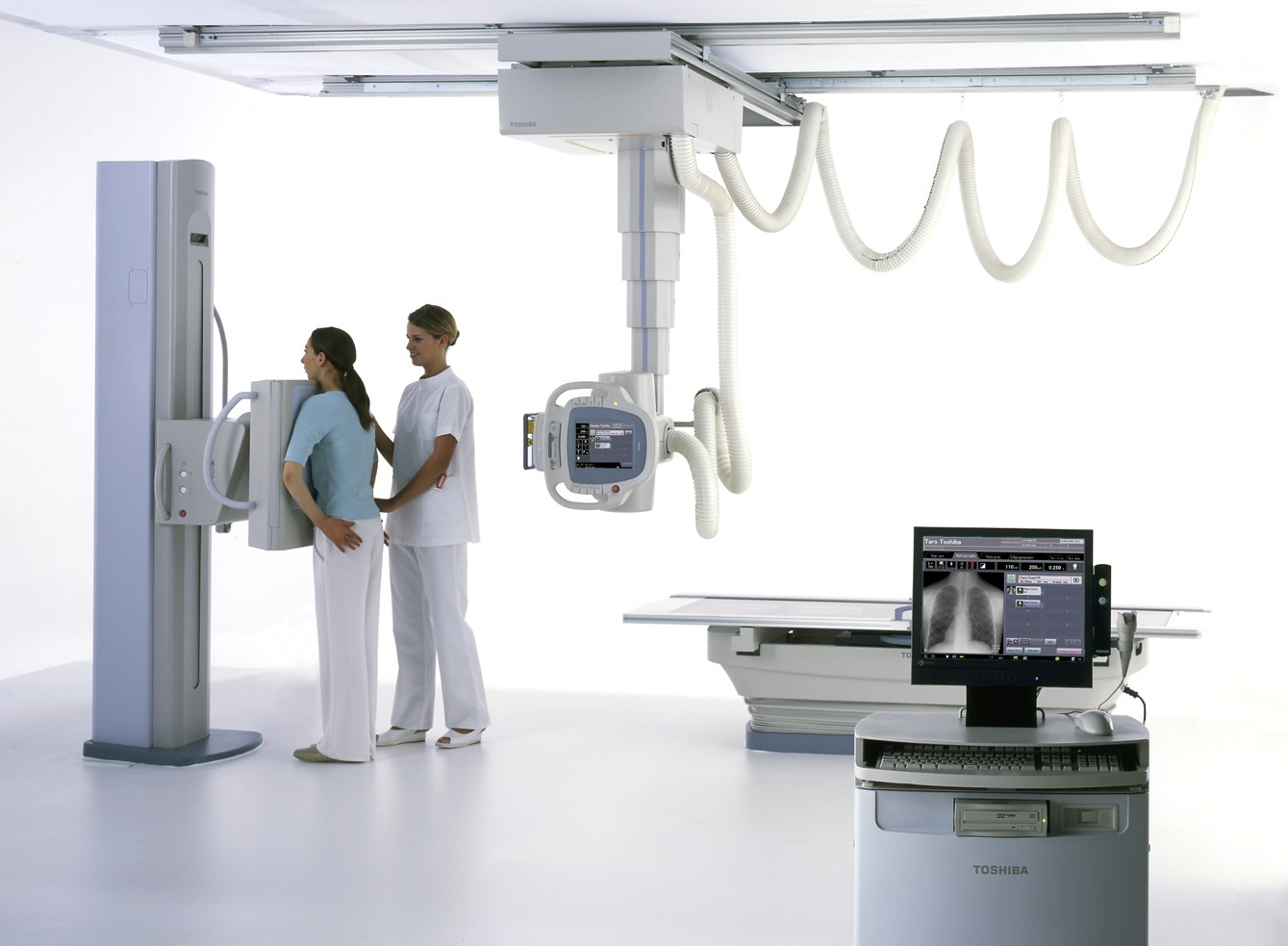
One of the oldest and most common types of medical imaging is x-ray imaging.
An x ray examination uses a machine to create a controlled beam of electromagnetic radiation. The beam goes through the body and hits a detector. Radiation is absorbed by various tissues in differing amounts, creating an image.
Their bones are well seen, as they have a higher X-ray absorption rate than many of the soft tissues around them.
What Are X Rays Used For?
Healthcare professionals commonly use X rays to examine:
- Broken or fractured bones
- Joint problems
- Chest conditions
- Lung abnormalities
- Certain infections
- Dental structures
- Some foreign objects
- Changes in bone alignment
Chest X rays are especially common. They can give information on the size of the lungs, heart, and some parts of the chest.
Advantages of X Rays
X rays are typically quick, and they are readily available. They can also be used to yield valuable information at relatively low cost to some advanced imaging techniques.
But regular X rays give only a little information on many soft tissues. They also employ ionizing radiation, and healthcare professionals question the necessity of the examination.
Computed Tomography
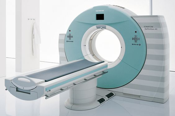
Computed tomography, or CT, is the use of X rays and computer-processing to generate detailed, cross sectional images of the body.
A CT scanner takes a series of measurements rather than just one image as the X ray source rotates around the patient. This information is fed into a computer which produces images called slices.
These slices can be displayed individually, or displayed in various planes and in some instances even three dimensional.
What Is a CT Scan Used For?
CT imaging can help evaluate:
- Head and brain injuries
- Internal bleeding
- Lung conditions
- Abdominal pain
- Kidney stones
- Bone fractures
- Blood vessels
- Certain cancers
- Complex traumatic injuries
In emergency medicine, exams can sometimes be performed on a rapid time frame, which is extremely useful with CT.
CT With Contrast
In some CT examinations, a contrast agent is given to help differentiate certain tissues, blood vessels or abnormalities.
Contrast can be injected into a vein or swallowed, depending on the exam.
Patients may have to give information about any previous reaction to the contrast, kidney or other medical conditions before taking the contrast.
Magnetic Resonance Imaging
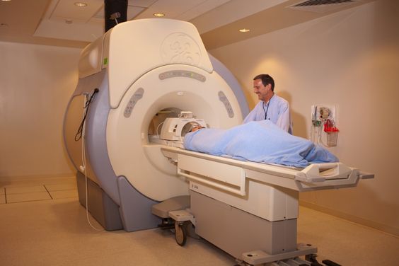
MRI (magnetic resonance imaging) generates detailed pictures with a strong magnetic field, radiofrequency energy and computer-processing.
MRIs do not use ionizing radiation, as do conventional X rays and CT scans.
MRI is particularly useful for examining soft tissues. It can generate a detailed image of the brain, spinal cord, muscles, ligaments, tendons, joints and numerous internal organs.
What Is an MRI Used For?
Doctors may recommend MRI to investigate:
- Brain abnormalities
- Spinal conditions
- Joint injuries
- Ligament and tendon damage
- Muscle injuries
- Certain tumors
- Abdominal conditions
- Pelvic conditions
- Blood vessels
MRI may be particularly useful for gathering soft tissue information that cannot be obtained from a conventional X ray.
What Happens During an MRI?
Patients generally lie on a table that is movable into the MRI scanner.
The examination may generate loud tapping, knocking or buzzing sounds. The patient will typically be asked to lie still, so the images are clear.
In some MRI studies, contrast is applied to highlight particular tissues and/or abnormalities.
Due to the powerful magnetic field, the MRI imaging team will ask patients to let them know about metal objects, implanted medical devices, or other pertinent materials before the exam.
Ultrasound Imaging
In ultrasound, high frequency sound waves are used to create images of structures within the body.
The ultrasound device, known as a transducer, is a hand-held device that sends sound waves through the body. These waves bounce off each other in different ways when they hit different tissues. The echoing waves go to the system that receives them and forms an image.
The use of ultrasound does not involve the use of ionizing radiation.
What Is Ultrasound Used For?
Ultrasound can help examine:
- Pregnancy
- Gallbladder
- Liver
- Kidneys
- Thyroid
- Blood vessels
- Muscles and tendons
- Certain soft tissue abnormalities
- The heart
Ultrasound may also be used to help doctors locate a needle to place, or to perform a biopsy.
Why Is Ultrasound Important?
A big benefit of ultrasound is that it can offer real-time imaging. Structures can be monitored in motion and blood flow can be detected by using special ultrasound techniques.
It can also be carried on a wheelchairs, making ultrasound valuable in the operating room, clinics and the emergency room.
Mammography
Mammography is a special type of X ray technology used to look at the breast tissue. It is a significant role in the breast cancer screening and diagnostic evaluation.
In a mammogram, the breast is placed and compressed between two imaging surfaces. Compression reduces the motion and can enhance image quality while using an appropriate radiation dose.
Screening and Diagnostic Mammography
Generally, screening mammography is an imaging performed in individuals who do not have a definite breast symptom as part of a screening program.
Diagnostic mammography might be suggested if someone experiences a breast symptom or if an area is identified during a screening exam that needs further evaluation.
Other imaging tests, such as ultrasound, may be used in addition to mammography depending on the situation.
Nuclear Medicine Imaging
Nuclear medicine gives information on how organs and tissues work, not just on their physical structure.
A small tracer of radioactive material is injected into the body. Tracer goes to the tissues based on their biological characteristics.
The radiation emitted by the tracer is captured by a special camera that forms images of the distribution of the tracer.
What Is Nuclear Medicine Used For?
Nuclear medicine examinations can help evaluate:
- Bone activity
- Thyroid function
- Heart function
- Kidney function
- Certain cancers
- Organ function
- Metabolic activity
This functional data can be used in addition to structural data such as from computed tomography (CT) or magnetic resonance imaging (MRI).
Positron Emission Tomography
Another type of nuclear medicine imaging is positron emission tomography (PET).
In PET, a radioactive substance is detected to demonstrate metabolic activity or biochemical changes in the body. A glucose related compound, which is capable of accumulating in tissues with increased metabolic activity, is commonly used.
CT is frequently used with PET, which results in PET/CT imaging. This combination offers functional and anatomical information.
What Is PET Imaging Used For?
PET can play an important role in:
- Cancer evaluation
- Cancer staging
- Treatment response assessment
- Certain neurological evaluations
- Some cardiac investigations
Information from PET can provide physicians with information about not only where an abnormality is located, but also how active certain tissues may be.
Medical Imaging in Cancer Diagnosis
Medical imaging has become an integral part of cancer treatments.
Imaging can assist physicians in finding an area that appears suspicious, measuring the size of the area, assessing adjacent tissues, checking for spread, and tracking changes as treatment progresses.
Various imaging modalities provide various kinds of information.
For instance, CT can offer detailed anatomical data about numerous regions of the body. Excellent soft tissue contrast can be achieved with MRI. PET can give information on metabolic activity.
In many instances, more than one imaging approach is required because none of the imaging techniques can provide the answer to all of the clinical questions.
It’s also crucial to note cancer is not always diagnosed by an imaging test. A biopsy or other test may be indicated if necessary, to make a definite diagnosis.
Medical Imaging for Emergency Care
Imaging is a significant part of EM for two reasons: Physicians are often in need of timely answers.
If the patient has sustained a severe injury, they may require imaging to determine if there is a fracture or internal bleeding. Brain imaging might be used in a person who suddenly has neurological symptoms to look for causes of the symptoms.
In emergency situations, CT imaging is often valuable due to its ability to generate detailed images in a timely manner.
Clinicians can also use ultrasound to assess some conditions at the bedside. Under certain circumstances, MRI might be used to add information when time and patient stability permit.
This decision will be made based on the patient’s condition and the problem suspected.
How Doctors Choose the Right Imaging Test
No single ideal imaging test for each of the symptoms. There are a number of factors that physicians consider before ordering a scan.
The Main Factors Include:
- The body part being examined
- The suspected condition
- The patient’s symptoms
- The urgency of the situation
- Age and overall health
- Previous imaging results
- Whether radiation is appropriate
- Whether contrast is needed
- The level of anatomical detail required
- Availability of the imaging technology
For example, an X ray may be required for a suspected broken arm and an MRI for suspected ligament injury.
That is why it is important for patients to not presume that the most sophisticated or costly scan is best.
The best test is the one that will yield the information to answer the particular clinical question.
What Happens Before a Diagnostic Scan?
Preparations vary according to type of examination. Other imaging tests do not require any special preparation. Others might need to fast, drink water, alter medications or take other advice.
Before the appointment, patients may be asked about:
- Current medications
- Allergies
- Previous reactions to contrast agents
- Pregnancy or possible pregnancy
- Implanted medical devices
- Kidney or other health concerns
- Previous imaging examinations
The instructions before the examination can help you to ensure that useful images are obtained during the examination.
What Happens During Medical Imaging?
The exact process differs for each technology.
An X ray can be made in just a few minutes. CT may also be fairly rapid (may require contrast and preparation).
MRI typically takes longer, and the patient must be still during the scan to get images.
In many cases, ultrasound will require the use of gel on the skin and the movement of a transducer over the area being examined.
Examinations with a nuclear medicine tool may be longer because the tracer needs to circulate around the body for some time before imaging is carried out.
The imaging team should be followed by patients during the study.
Are Medical Imaging Scans Safe?
In today’s medical practice, medical imaging is a universal technique, while the safety depends on the technique.
X rays and CT involve the use of ionizing radiation. The benefit of the examination is weighed against potential radiation exposure by healthcare professionals.
Despite not using ionizing radiation, MRI does have specific safety requirements due to the powerful magnetic field.
Unlike x-rays, ultrasound does not involve the use of ionizing radiation, and it is used in many clinical uses.
During the course of nuclear medicine, the person receives a small dose of radiation from the radioactive tracer.
The appropriate imaging test depends on the clinical situation. Patients should tell their healthcare provider about pregnancy, implanted devices, allergies, medications, and other relevant concerns before an examination.
What Is Contrast Material in Medical Imaging?
Certain tissues are enhanced with contrast agents, which aid in their better visualization during an imaging examination.
It does not just lighten all pictures. Rather, it can enhance the contrast between distinct tissues, vessels or abnormalities.
Different imaging technology requires different contrast agents.
For instance, in some exams, CT could be given an iodine contrast dye, and in certain cases, MRI might be given a gadolinium based contrast dye.
Not all scans require contrast.
It is administered at the discretion of healthcare professionals and is determined by their assessment of whether it has value to provide added information for the patient.
Benefits of Medical Imaging
Medical imaging has transformed modern diagnosis.
Some major benefits include:
- Detecting disease earlier
- Identifying injuries without exploratory surgery
- Supporting accurate diagnosis
- Monitoring treatment
- Guiding procedures
- Assessing disease progression
- Providing anatomical information
- Showing certain biological processes
Imaging also can eliminate doubt. A doctor could have a number of different diagnoses for a patient’s symptoms. A good choice of scan can give information that can help eliminate those options.
Limitations of Medical Imaging
Although useful, medical imaging has several drawbacks.
An image might show an abnormality but not tell what is causing it. There may be some findings that are not specific. Sometimes the scan is normal even if the patient has symptoms.
Imaging also may reveal incidental findings. They are unusual conditions which may or may not be related to the reason for the scan.
Sometimes more testing may be required.
Doctors, therefore, draw conclusions based on the image in the context of their overall clinical situation, and not as a stand-alone conclusion.
The Role of Radiologists
Radiologists are doctors focused on reading medical images and aiding in the diagnosis of diseases and injuries by using imaging.
Once examined, a radiologist looks at the images and creates a report detailing any pertinent results of the examination.
Normal findings, abnormalities, measurements, comparisons with previous exams and recommendations for further imaging (if appropriate) may be noted in the report.
The referring health care provider then reviews the radiology report as well as the symptoms, examination, lab findings and other information.
This is a collaborative process rather than the process itself, which is an important component of diagnostic medicine.
Artificial Intelligence and the Future of Medical Imaging
Artificial intelligence is becoming a part of medical imaging workflow more and more.
AI systems can help the tasks like image analysis, prioritization of potentially urgent examinations, measurements, image reconstruction, and detection of particular patterns.
But AI is not a replacement for trained healthcare providers!
The most useful function of AI is likely to be to assist physicians in interpreting vast quantities of imaging data quickly and pinpointing areas that might need further investigation.
As these technologies evolve, medical imaging could also be quicker, more individualized and more interconnected with other clinical information.
Medical Imaging and Personalized Healthcare
Diagnostic imaging is not just about seeing the picture on the screen; it is moving on.
Present imaging techniques can provide structural, functional and quantitative information. Imaging can complement laboratory and genetic information, along with patient histories, to aid in individual treatment decisions.
As an example, imaging can help assess tumor features and response to treatment over time in cancer treatment.
This change is significant because increasingly health care is driven by an understanding of the behavior of a particular patient’s disease, not just a diagnosis.
Key Takeaway
Medical imaging is used to provide medical professionals with the ability to see inside the human body and get information that could not be gained from a physical exam. There are many uses of x-rays for bones and chest imaging, and CT can provide detailed cross sectional imaging, MRI can provide good imaging of soft tissue, ultrasound can provide real time imaging without the use of ionizing radiation, and nuclear medicine can give information on biological activity.
The appropriate scan to use is not only determined by the most cutting-edge technology but by the clinical question at hand.
Further advancement of imaging technology will allow for further enhancements to image quality, workflow productivity, artificial intelligence, and quantitative analysis, making diagnostic imaging more useful. The most crucial step is to discuss with the health care team the purpose, benefits, risks, and preparation needs for a recommended examination for any patient.
Medical imaging is most effective when it is used in conjunction with clinical expertise and patient specific information, and the quality of the images.
Frequently Asked Questions About Medical Imaging
What is medical imaging used for?
Medical imaging is imaging of the body’s structures and processes. Imaging is used in medicine to identify disease, diagnose injury, assess symptoms, plan procedures, plan treatment and monitor changes over time.
What are the four main types of medical imaging?
There are four main types: X ray, CT, MRI and ultrasound. Other imaging technologies that are important to the doctor include nuclear medicine and PET. When doctors need information about organ function or metabolic activity, these imaging technologies are important.
Which medical imaging method is best?
There is no universally best imaging method. The test used will be different for different parts of the body, for different suspected conditions, for different clinical questions, for different patient factors and for different urgency.
Does medical imaging use radiation?
Imaging technologies that utilize ionizing radiation include X ray, computed tomography (CT), mammography and nuclear medicine. MRI and ultrasound don’t employ ionizing radiation
Is MRI safer than CT?
There are safety concerns with MRI and CT. MRI is not radioactive, but has some limitations. Although CT involves the use of ionizing radiation, it can also yield useful information relatively rapidly, particularly in emergency situations. This depends on medical need and should be determined accordingly.
How long does a medical imaging scan take?
The time frame is very variable. Some X rays can take a matter of minutes, and MRI may take a lot longer. Nuclear medicine procedures may also be longer since imaging can be performed after the tracer has spread throughout the body.
Can medical imaging detect cancer?
Medical imaging may detect abnormal masses, abnormalities and changes that could be cancerous. But imaging is not always enough to make a definitive diagnosis of cancer. Further tests such as a biopsy might be needed.