









Doctors use nuclear medicine imaging to see how the cells inside the body are functioning. The specialized field of radiology looks at how your organs ACTUALLY function in real time, as opposed to a series of static snapshots of bones and organs. While patients may be anxious when their doctors ask them to have a nuclear scan, they are one of the safest and least invasive diagnostic imaging tests available in modern medicine.
This complete guide explains the uses, methods, and major categories of nuclear imaging tests; the significance of the scans; and the safety and side-effects you can expect.
What Is Nuclear Medicine Imaging?
Nuclear medicine imaging is a specialized form of medical radiology that involves the use of small amounts of radioactive substances (radioisotopes or radiopharmaceuticals, also known as radiotracers) for diagnostic, evaluation, and therapeutic purposes in the treatment of a variety of diseases.
Traditional x-rays, magnetic resonance imaging (MRI) and computed tomography (CT) scans examine the anatomy while nuclear medicine looks at the function and metabolic activity of cells. This is an important distinction because doctors are able to detect even the smallest changes in these physiological processes before it shows up on standard imaging devices.
How Radiotracers Work in the Body
Radiotracers are molecules that are tightly attached to a very small amount of radioactive material. The choice of the radiotracer depends on the organ or tissue under evaluation.
- Administration: Medical staff inject the radiotracer into a vein, give it as an oral liquid or capsule, or ask you to inhale it as a gas.
- Targeting: The tracer naturally travels to specific organs or biological pathways. For instance, thyroid tissue quickly absorbs iodine, while rapidly growing tumor cells consume high amounts of glucose.
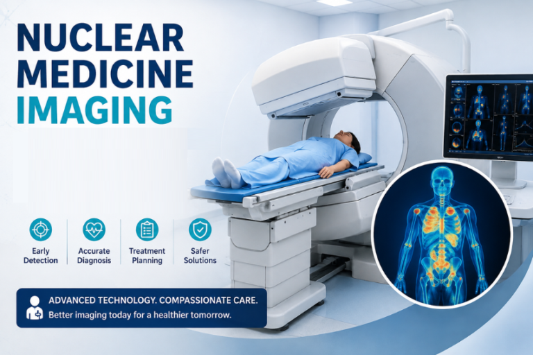
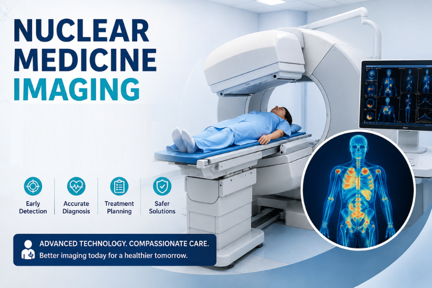
- Detection: As the radiotracer collects in specific tissues, it emits gamma rays. A specialized computer camera detects these emissions and translates them into detailed 3D pictures.
How Nuclear Medicine Scans Differ from Standard X-rays
Understanding the core differences between nuclear imaging and conventional radiology helps demystify the scanning process.
| Feature | Standard X-Rays / CT Scans | Nuclear Medicine Imaging |
|---|---|---|
| Primary Focus | Physical anatomy and structure | Biological function and cellular metabolism |
| Radiation Source | External machine passes rays through the body | Internal radiotracer emits signals from inside the body |
| Early Detection Capabilities | Detects changes after structural damage occurs | Identifies disease at the cellular level before structural changes occur |
| Primary Application | Fractures, lung infections, structural tissue damage | Early cancer detection, heart tissue viability, organ performance |
Major Types of Nuclear Medicine Imaging Tests
There are a number of special nuclear scans used in healthcare that can show different parts of the human body working.
Positron Emission Tomography (PET)
PET scans detect the high-energy gamma photons emitted by radioactive substances, or radiotracers, that are usually based on fluorodeoxyglucose (FDG), a substance that looks like a natural sugar. Cancer cells have a high level of glucose uptake making them easily visible as “hot spots” on the final image, because they grow quickly and require more glucose than healthy cells. PET scanners are often used in combination with conventional CT scanning (PET-CT), to identify the exact location of abnormal cells.
Single-Photon Emission Computed Tomography (SPECT)
SPECT is a method that generate cross sectional 3D images of internal organs by rotating a gamma camera. SPECT is similar to a PET scan but it employs other radiotracers that emit one gamma ray. SPECT is crucial to cardiologic tissue evaluation and plays a significant role in creating detailed maps of blood flow through the heart and brain, which is critical in stroke evaluations.
Bone Scans
A nuclear bone scan assesses the metabolism of bone throughout the body. Radiotracers attach rapidly to areas where bone is actively repaired or new bone cells are being created. This test is very good at detecting micro-fractures, bone infections (osteomyelitis), primary bone tumors, and spread of cancer to the bones from other parts of the body.
Thyroid Scans and Uptake Studies
Iodine is essential for the thyroid gland to create the metabolic hormones that are vital for the body. In a thyroid uptake test, a small amount of radioactive iodine is given as a pill or liquid and is swallowed. The imaging technician takes measurements of the amount of iodine taken up by the thyroid over a time period. Most of the time, high absorption rates suggest hyperthyroidism or Graves’ disease, while low absorption rates can suggest thyroid inflammation or hypothyroidism.
Cardiac Stress Tests (Myocardial Perfusion Imaging)
During a nuclear cardiac stress test, blood flow in your coronary arteries is evaluated while you are at rest and after physical stress. Enjoy a walk on the treadmill or receive a drug that mimics exercise while a radiotracer is injected into you. The scans give a visual representation of where the blood is not flowing as well, which shows blockages in the arteries or damaged heart muscle after a heart attack.
HIDA Scans (Cholescintigraphy)
HIDA scan measures the activity of the liver, gall bladder and bile ducts. The camera follows the formation of bile in your liver and its movement from that area to your gallbladder and small intestine following the injection of a tracer. A HIDA scan may be ordered to help diagnose acute gallbladder inflammation, obstructions of the biliary tract, and leaks after surgery.
Renal (Kidney) Scans
Renal scintigraphy helps to assess blood flow to the kidneys, individual kidney function and flow of urine. This scan is used by specialists to assess kidney transplants, as well as to diagnose high blood pressure as a result of narrowing in the renal arteries, and to determine if there are any blockages in the urine flow from the kidneys, in paediatric and adult patients.
Primary Uses of Nuclear Medicine Diagnostics
Nuclear medicine plays a transformative role across multiple medical specialties, offering diagnostic clarity that helps teams create precise, personalized treatment plans.
Oncology Applications
Cancer management relies extensively on nuclear imaging. Physicians utilize these tests to:
- Identify primary tumors at an early phase.
- Determine whether cancer cells have spread to nearby lymph nodes or distant organs.
- Evaluate how effectively a tumor responds to chemotherapy or radiation therapy.
- Detect recurring cancer long before structural scans show visible tissue changes.
Cardiovascular Applications
Cardiologists use nuclear scans to make crucial decisions about surgical interventions and long-term care. Key uses include:
- Evaluating blood circulation through heart walls.
- Determining the extent of permanent heart tissue damage after a heart attack.
- Checking the success of bypass surgeries or stent placements.
Neurological Applications
The human brain consumes vast amounts of energy, making functional nuclear imaging ideal for complex brain assessments:
- Differentiating Alzheimer’s disease from other causes of dementia.
- Locating precise brain regions responsible for epileptic seizures prior to surgery.
- Evaluating blood flow patterns in individuals who have suffered strokes or traumatic brain injuries.
- Assessing dopamine pathways to aid in diagnosing Parkinson’s disease.
Bone and Joint Applications
Orthopedic specialists use bone scintigraphy to discover subtle musculoskeletal issues that standard X-rays routinely miss:
- Uncovering stress fractures in athletes.
- Detecting subtle prosthetic joint infections or loosening.
- Investigating unexplained, persistent bone pain.
What to Expect During Your Test
Understanding the standard steps of a nuclear medicine procedure helps relieve anxiety and ensures smooth, accurate diagnostic results.
Step 1: Pre-Test Preparation
Preparation steps vary based on the exact scan ordered:
- Fasting: PET scans typically require complete fasting for four to six hours beforehand to keep blood glucose low.
- Medication Adjustments: You may need to temporarily stop taking specific heart medications or thyroid supplements.
- Hydration: Many scans encourage drinking plenty of water before arriving to speed up tracer distribution.
- Clothing: Wear comfortable clothing without metal zippers, snaps, or belts.
Step 2: Radiotracer Administration
Upon arrival, a specially trained nuclear medicine technologist injects the radio-tracer into the IV line, swallows the dose or uses a special inhalation mask. The injection is the same as a regular blood draw or IV placement.
Step 3: The Circulation Period
You will lie very still as the radiotracer passes through your body and accumulates in the organ of interest. The waiting time varies based on the test, from 30 minutes to a few hours. In PET scans, you will lie still in an enclosed room; talking or movement will alter the distribution of the tracer to undesirable muscles.
Step 4: The Scanning Process
During the scan you rest on the padded examination table facing down. The large gamma camera or PET scanner glides around you, close to your body. Unlike older MRI machines, it isn’t noisy and doesn’t require small dark tubes. It is very important that you remain absolutely still while undergoing imaging passes, typically lasting 20-45 minutes.
Step 5: Post-Test Recovery
The technologist, after checking the quality of the image, can then return you to your normal daily routine, work and driving.
Safety Facts, Risks, and Side Effects
Safety concerns regarding radiation exposure remain a top question for patients. Nuclear medicine tests are safe, highly controlled diagnostic procedures designed to minimize risks.
Radiation Exposure Levels
Low doses of short-lived radiopharmaceuticals are employed for nuclear medicine imaging. The amount of radiation received during a normal scan is sometimes comparable to the normal background radiation you might receive in a few months or years from the environment.
The radioactive materials used will decay quite quickly from the body by natural physical decay, so that the majority of radiation will be expelled within a few hours to days.
Common vs. Rare Side Effects
Actual side effects remain exceptionally rare because the body receives only minute traces of active pharmaceutical compounds.
- Minor, Short-Term Effects: Mild redness or temporary soreness at the injection site.
- Uncommon Reactions: Mild metallic taste in the mouth or brief skin flushing.
- Rare Allergic Reactions: Skin rashes, mild itching, or slight fever. Serious allergic reactions occur far less frequently than those associated with standard CT contrast dyes.
Patient Safety Guidelines
To minimize exposure risks for others, keep these simple, practical precautions in mind:
- Hydration: Drink plenty of water throughout the day following your test to accelerate flushing the tracer through your kidneys.
- Pregnancy and Breastfeeding: Always inform your doctor and technologist if you are pregnant, planning to become pregnant, or currently nursing. Certain radiotracers can cross the placenta or pass into breast milk, requiring alternative test options or brief pump-and-discard schedules.
- Close Contact Precautions: For a short window, usually 6 to 24 hours depending on the tracer used, providers may suggest maintaining a safe distance from infants and pregnant women as a standard safety precaution.
Nuclear Medicine vs. Standard Imaging Modalities
Choosing the proper imaging tool depends entirely on what your medical provider needs to observe.
- CT Scans: Use targeted X-rays to generate sharp anatomical cross-sections. They excel at identifying structural injuries, bone fractures, and physical masses.
- MRIs: Rely on radio waves and strong magnetic fields to image soft tissue details, like ligaments, brain tissue, and spinal discs, without ionization.
- Nuclear Medicine: Measures cellular metabolism, organ blood flow, and biochemical changes.
Combining functional nuclear scans with anatomical CT or MRI scans gives medical teams an exceptionally detailed view of internal health issues.
Final Thoughts
Nuclear medicine imaging test provides helpful information that will help your doctor understand how the body works on a cellular level. These functional tests enable physicians to identify changes to the body at an early stage, improving the ability to identify complex conditions in more timely fashion; to properly assess treatments; and create a targeted recovery plan.
When your doctor suggests a nuclear medicine test, know that these safe, non-invasive tests can provide strong answers to help make informed decisions about your health.